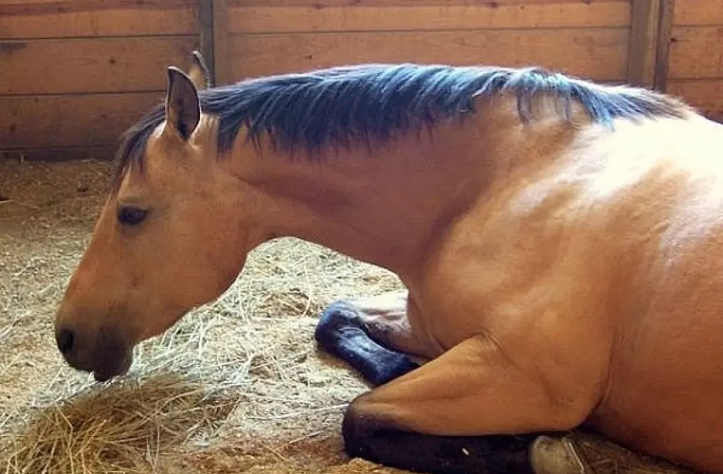
Colic, or abdominal pain, is a common ailment in horses. More than 70 causes can trigger colic, including gas distention, food impactions, intestinal tract spasms, and intestinal displacement or twists. One of the more exotic forms is colic caused by enteroliths, or stone-like formations that form in a horse’s digestive tract.
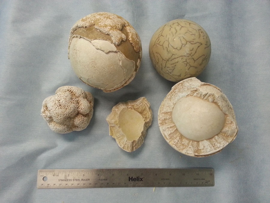
Enterolith stones are made up of minerals, such as magnesium ammonium phosphate salts. These minerals can build up around an object that a horse eats but does not digest, such as a small chunk of wood, pebble, wire, twine, or other foreign object. These masses can become quite large, sometimes weighing in at 10 to 15 pounds or more.
 Sometimes horses develop one large stone, while others chronically develop multiple stones. The stones form in the large colon of the horses and cause a problem once they move to areas of the intestine that have smaller diameters, such as the transverse colon or the small colon.
Smaller enteroliths might pass with feces, but if horses produce smaller stones, larger ones might remain. Mild chronic or intermittent colic can plague horses with a single stone that has not grown large enough to cause a total impaction or has not moved to a narrower area. These horses might also suffer from weight loss, diarrhea, or lethargy.
There are several risk factors for enterolith development. Geography is one. Certain areas like the southwestern U.S. produce many more cases than others, particularly California and Arizona. In California, enteroliths are the leading cause of surgical colic cases. The mineral composition of the soil and feed material grown there are suspected culprits. The risk does not seem as high in the Northwest, but approximately five cases are seen at the WSU Veterinary Teaching Hospital each year.
“Even horses that travel to Arizona or California for shows or other events can develop enteroliths while visiting,” said Julie Cary, DVM, MS, Dipl. ACVS, clinical instructor of equine surgery and emergency care at WSU.
Enteroliths also seem more common in horses fed a diet of more than 50% alfalfa hay. They are more commonly found in adult horses between 10 and 15 years of age, in female horses, and in the Arabian horse breed, although the problem can occur in horses of any breed.
If a horse is suspected to have enteroliths, the best way to confirm a diagnosis, other than exploratory surgery, is to take abdominal radiographs or X rays. Large veterinary hospitals or clinics usually have a radiograph generator powerful enough to produce a picture of a horse’s abdomen.
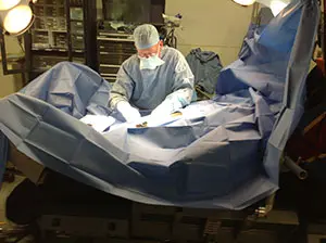
If surgery is required, most horses have a very good outcome unless a portion of the intestine was badly damaged or ruptured. After surgery, horses generally need three months to recover before returning to full exercise. Owners should continue to monitor them for colic. Stall rest is required for about a month before being placed back to pasture.
Because enteroliths are uncommon in the Pacific Northwest, the faculty at the Veterinary Teaching Hospital does not recommend specific preventive measures for most horses. However, if your horse has been diagnosed with an enterolith in the past, the following measures may help prevent recurrence:
- Decrease alfalfa hay in the diet and feed good quality grass hay instead.
- Don’t feed bran because it contains very high levels of phosphorus.
- Add a cup of vinegar a day to your horse’s diet to help decrease the pH inside the intestine.
- Provide your horse with frequent access to pasture. Continual grazing will help to encourage regular movement of feed material through the intestines.
- Ask your veterinarian about including occasional doses of psyllium in your horse’s diet to increase bulk movement through the intestines.
- Provide regular, consistent exercise for your horse.


